EXCLUSIVE: Natural News releases post-vaccine clot ICP-MS elemental analysis results, comparing clots to human blood … findings reveal these clots are NOT “blood” clots
08/17/2022 / By Mike Adams

We are now releasing ICP-MS lab test results that compare the elemental composition of human blood to the elemental composition of a clot sample taken from the body of a person who received a covid vaccination and then subsequently died. This clot was provided by embalmer Richard Hirschman, and these clots are being widely reported in the bodies of people who have “died suddenly” in the weeks or months after receiving one or more covid vaccinations.
According to rigorous analysis based on excess death data — summarized nicely by Steve Kirsh at Substack — there are currently around 10,000 people dying each day from covid vaccines. Anywhere from 5 to 12 million fatalities have likely occurred worldwide so far, and with these self-assembling clots continuing to gain size and mass inside the bodies of those who have received the mRNA experimental medicine injections, it is certain that many people who have not yet died from the vaccines will experience death in the coming months and years.
Kirsch has roughly estimated that 1 person is currently dead for every 1,000 doses of covid vaccines that are administered. This number will almost certainly increase with time, as the clots that are causing so much death appear to be continuing to “grow” (self-assemble) inside the blood vessels and arteries of vaccine victims. Thus, the final toll of covid vaccines will only be experienced over a period of several years and could be orders of magnitude higher, potentially 1 in 100 or even 1 in 10, although we will have to watch excess deaths carefully over the next several years to know where this post-vaccine death phenomenon levels out.
So far, over 12 billion covid vaccine doses have been administered worldwide. Over 600 million doses have been administered in the United States, and Kirsch estimates that 600,000 Americans have likely already been killed by covid vaccines in the USA alone. (That’s about 12 times higher than the total casualties of US soldiers in the Vietnam War, for comparison.)
Here’s a photo that I took of one of these clots, under a lab microscope:

Pursuing the mystery of the post-vaccine clots
Dr. Jane Ruby has been one of the researchers at the forefront of attempting to determine the composition of these clots as well as their mechanism of action in causing fatalities in victims. Dr. Ruby connected us with Hirschman and helped arrange for the clot samples which we have tested via ICP-MS in our ISO-accredited, 17025 approved laboratory which specializes in food and water analysis.
In full disclosure, our laboratory is accredited, audited, inspected and validated for ICP-MS testing in food and water samples, as well as other areas such as cannabinoid quantitation analysis in hemp extract samples. However, the accreditation scope of our lab does not specifically encompass human biological samples, as we do not offer such testing to the public. Nevertheless, we routinely test dog food and cat food samples which are, of course, composed of animal flesh and ground blood vessels, meat tissue, cartilage and other animal-derived biological structures, and we are using the exact same sample preparation, digestion, analysis and reporting methods for post-vaccine clot samples. We also routinely test beef, poultry, fish and other meat samples. Thus, we are highly confident in the accuracy of these results. Furthermore, we did not see any failures during the sample prep process. The entire clot was dissolved in nitric acid, meaning its elements went into solution and were able to be analyzed via ICP-MS.
Here’s a photo of some of the clots found in the body of the deceased:

These ICP-MS tests were conducted on June 23 of this year. We have delayed public release of the results in order to allow time to share these numbers with colleagues and to invite feedback from others. These PDFs have also been shared privately with Dr. Jane Ruby and others. No one with expertise in this field has indicated any apparent problems or concerns about this analysis. If anything, the ICP-MS analysis is rather straightforward: Samples are “digested” into nitric acid, this acid is nebulized into a liquid stream which goes through a plasma torch, gets ionized and then directed through a quadrupole assembly that sorts the elements by their mass-to-charge ratios. Each individual element is scanned and counted on a PMT (Photo Multiplier Tube) which translates individual elements into electrical current that can be accurately counted. These results are mapped against external standards which are NIST traceable to provide very accurate calibration curves, which means the quantitation data are extremely reliable.
We used 0.4528 grams of the clot as the sample mass in this case:

For a primer on ICP-MS and why it is so accurate, see this NIH article.

ICP-MS analysis results reveal that these clots are not made of blood – they are not “blood clots”
Although we intend to conduct more tests on clots and blood samples, the data we see so far make it clear that these clots are not “blood clots.” They are not simply made of congealed blood.
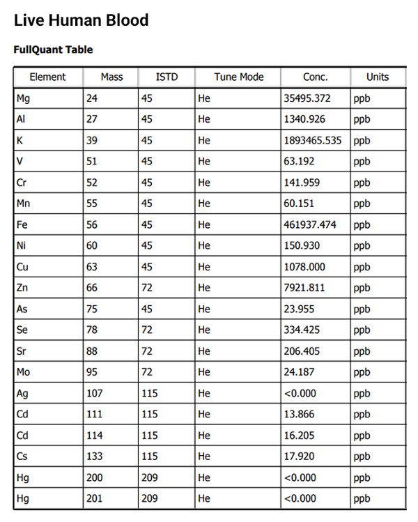
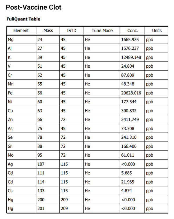
How do we know this? Because the elemental ratios and densities are vastly different. Consider the following comparison chart, based on our ICP-MS results (see full results below), and notice the stark differences between the elemental concentrations in blood vs. clot among nutritive “marker” elements such as iron and magnesium:
| Element | Blood Results | Clot Results |
| Mg (magnesium) | 35 ppm | 1.7 ppm |
| K (potassium) | 1893 ppm | 12.5 ppm |
| Fe (iron) | 462 ppm | 20.6 ppm |
| Zn (zinc) | 7.9 ppm | 2.4 ppm |
| Cl (chlorine) | 930,000 ppm | 290,000 ppm |
| P (phosphorous) | 1130 ppm | 4900 ppm |
As you can see, the post-vaccine clot sample only contains 4.4% of the iron that would be seen in human blood. This alone is confirmation that this clots is not a “blood clot.” In addition, note the near-total lack of potassium (K) in the clot sample. The clot contains less than 0.6% of the potassium as human blood. It’s a similar story with magnesium, too.

Several electrically conductive elements were higher in the clot
In addition to the nutritive elements shown above, we noticed a peculiar pattern among electrically conductive elements such as sodium (Na), aluminum (Al) and tin (Sn). For the following table, please note that the tin and sodium results come from a separate “semiquant” report which is less accurate than the “fullquant” analysis used for all the other elements shown here. In essence, the semiquant numbers are accurate in terms of relative concentrations from one sample to the next, but they are not compared to calibrated external samples, so the actual (absolute) concentration reported does not have the confidence interval of the fullquant results:
| Element | Blood Results | Clot Results |
| Na (sodium) | 1050 ppm* | 1500 ppm* |
| Sn (tin) | 163 ppb* | 942 ppb* |
| Al (aluminum) | 1.3 ppm | 1.6 ppm |
* = SemiQuant results, not FullQuant
With sodium being nearly 50% higher in the clot, and tin showing an increase of 588%, we can only conclude that the self-assembling clot is, in effect, “harvesting” or concentrating certain elements from circulating blood as clot assembly is taking place. It is noteworthy that many of these elements are conductive. Aluminum, for example, is the most common alternative to copper for use in electrical wiring. Sodium is an alkali metal that is highly conductive, and tin is used as the primary component in solder alloys used to manufacture or repair circuit boards.
You can see the numbers on elemental conductivity at this electrical conductivity reference table from Angstrom Sciences.
One conclusion is inescapable: The clot is almost entirely lacking key marker elements that would be present in human blood (such as iron and potassium) yet shows significantly higher concentrations of elements that are used in electronics and circuitry.
We invite the reader to draw your own conclusion of the explanation behind that, merely noting that the patents of Dr. Charles Lieber may be of special interest.
This analysis, notably, does not answer any question of whether these clots are “alive” or dead (like hair and nails). My own professional opinion is that these clots are not living structures. They appear to be self-assembling dead biostructures, from what we can see so far. But that’s just an initial assessment and may change with additional observations or findings. Prions, for example, are self-assembling but non-living biostructures too. They are essentially mis-folded proteins that spread throughout the brain (or other regions), causing morphological alterations that nullify both the normal structure and function of neurological cells. Something does not have to be alive in order to be self-assembling. Even viruses, as described by traditional virology, are dead structures which are nevertheless self-assembling and can “grow” in size and mass in terms of their aggregate population.
The following microscopy picture, taken at our lab at around 1500 x magnification, shows what appears to be a repeating structure on a wire-looking protrusion from one of these clots. In case you were wondering, is not a human hair. It is connected to the clot:


See the ICP-MS results for yourself
For those not familiar with the units being reported here:
ppb = parts per billion
ppm = parts per million
1,000 ppb = 1 ppm (because the metric system)
The units used by the instrument are mass over volume (m/v) and the “mass” is technically mass-to-charge ratio (m/z).
Here’s a screen shot of a section from the PDF report of the ICP-MS results for live human blood:

You can also download the full PDF document for the blood analysis here.
And here’s the screen shot of the results from the clot analysis, showing ICP-MS analysis for the post-vaccine clot:

Finally, you can download the full PDF document of the ICP-MS analysis for the clot here.
Share these results and keep asking questions… more analysis yet to come
Feel free to share these results, incorporate them into your own videos or podcasts, and offer your own explanations for what might explain this apparent anomaly. Please give credit to NaturalNews.com as the source, as we conducted this exclusive analysis in order to help resolve the mystery of these clots that seem to be killing a large number of people.
We welcome any feedback on these results, including corrections if any errors are found.
We also encourage other labs to replicate these tests for yourself and publicly publish your findings as we have done here.
More analysis results are coming shortly, including additional microscopy images.
Join the Natural News email newsletter (free) to be alerted via email as we announce and release new lab science findings.
Submit a correction >>
Tagged Under:
This article may contain statements that reflect the opinion of the author
RECENT NEWS & ARTICLES
BadMedicine.News is a fact-based public education website published by BadMedicine News Features, LLC.
All content copyright © 2019 by BadMedicine News Features, LLC.
Contact Us with Tips or Corrections
All trademarks, registered trademarks and servicemarks mentioned on this site are the property of their respective owners.


